
What to expect
Your PET/CT scan, step-by-step
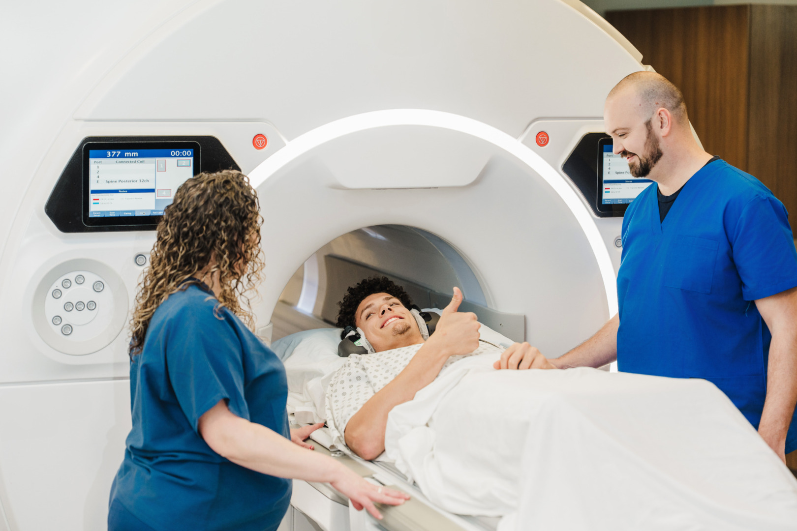
From your first call to your final results, Shields is here for you. Our PET/CT team walks you through preparation, care, and next steps—bringing advanced imaging and compassion together for you.
Scroll


Find a Shields Location
Search locations near you with details on services, hours, and technology.
A Patient’s Perspective